Introduction
Ultrasound imaging is a widely used diagnostic tool that employs sound waves to create images of internal organs and tissues. During an ultrasound examination, a transducer transmits sound waves into the body. These waves bounce back (echo) depending on the density of the tissue they encounter. The reflected sound waves are then converted into a visual representation on the screen, allowing doctors to assess the structure and function of various organs. One term you might encounter in your ultrasound report is hypoechoic. But what exactly does it mean, and why is it significant? Let’s delve into the world of ultrasound terminology and explore the implications of a hypoechoic finding.
Decoding: Less Echo, More Density
The term “hypoechoic” literally translates to “less echo.” In the context of ultrasound, it signifies that a particular area within the scanned tissue reflects fewer sound waves compared to the surrounding healthy tissue. This typically indicates a region of increased density or solidity.
Here’s a breakdown of the key terms:
- Echo: The reflection of sound waves.
- Hypo: Meaning “less than normal.”
- Echoic: Relating to the reflection of sound waves.
Therefore, a hypoechoic area appears darker on an ultrasound image because it absorbs more sound waves and generates fewer echoes.
Understanding Echogenicity: A Spectrum of Echoes
Ultrasound images depict tissues based on their echogenicity, which refers to the ability of a tissue to reflect sound waves. Here’s a spectrum of echogenicity you might encounter:
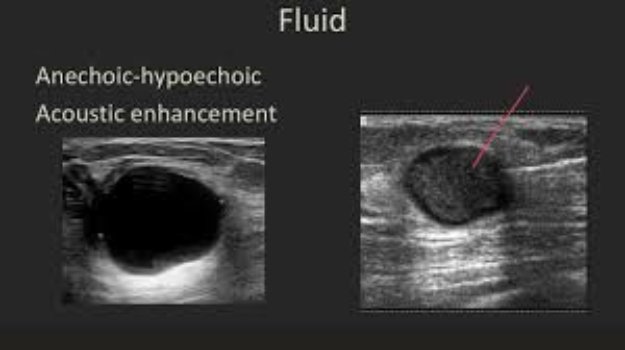
- Anechoic: Appears black on the image, signifying structures with minimal sound wave reflection, often fluid-filled cysts.
- Hypoechoic: Appears darker than surrounding tissue, indicating increased density.
- Isoechoic: Appears similar in echogenicity to surrounding tissue, considered normal.
- Hyperechoic: Appears brighter than surrounding tissue, suggesting structures with high sound wave reflection, like air or fat.
Common Organs Examined with Ultrasound
Ultrasound is a versatile imaging technique used to examine various organs and tissues throughout the body. Some frequent applications include:
- Liver: To assess for abnormalities like cysts, tumors, or fatty infiltration.
- Kidneys: To evaluate for kidney stones, cysts, or blockages.
- Thyroid gland: To detect nodules or masses within the thyroid.
- Uterus and ovaries: To examine for fibroids, cysts, or monitor pregnancy.
- Breast: To screen for potential breast lumps or masses.
When Does Hypoechoic Become a Concern?
While it finding on an ultrasound can be unsettling, it doesn’t necessarily signify a serious condition. Here’s why:
- Normal Variations: Certain tissues naturally exhibit hypoechogenicity. For instance, the liver appears darker than the kidneys on an ultrasound due to its denser nature.
- Benign Lesions: Non-cancerous growths like cysts or lipomas can also appear hypoechoic.
- Abnormal Growth: Area might represent a tumor or mass, which could be benign or malignant (cancerous).
- Inflammation: Inflamed tissues can sometimes appear denser and hypoechoic on ultrasound.
Importance of Additional Testing
An isolated hypoechoic finding on ultrasound is usually not enough to confirm a diagnosis. Further evaluation might be necessary depending on the specific organ involved, the patient’s medical history, and other factors. Here are some additional tests that might be employed:
- Biopsy: A small tissue sample is extracted for microscopic examination to determine if the tissue is cancerous.
- CT Scan: Provides detailed cross-sectional images using X-rays to offer a clearer picture of the area.
- MRI Scan: Utilizes magnetic fields and radio waves to create detailed images of organs and soft tissues.
FAQs
Here are some frequently asked questions regarding hypoechoic findings on ultrasound:
Should I be worried about finding?
Not necessarily. A hypoechoic finding can have various explanations, and additional tests are often required for a definitive diagnosis. Discuss your concerns with your doctor.
What determines the next steps after finding?
The course of action depends on several factors, including the organ involved, other ultrasound findings, and your medical history. Your doctor will guide you through the necessary follow-up steps.
Can a hypoechoic mass always be cancerous?
No. Many hypoechoic masses are benign. However, a biopsy might be needed to confirm the nature of the mass definitively.
Conclusion
Ultrasound imaging offers a valuable window into the inner workings of our bodies. While encountering a hypoechoic finding on your ultrasound report can be a source of worry, understanding its meaning empowers you to participate actively in your healthcare journey. Remember, it finding doesn’t automatically translate to a serious condition. It simply indicates an area of increased density that warrants further investigation. By working collaboratively with your doctor, you can navigate the next steps effectively. Additional tests might be recommended to reach a definitive diagnosis. Early detection and prompt intervention are crucial for managing any potential health concerns.
Ultimately, knowledge is power. This article has equipped you with the essential understanding of hypoechoic findings on ultrasound. Don’t hesitate to discuss any questions or concerns you might have with your doctor. They are your partner in navigating your health journey and ensuring optimal well-being.